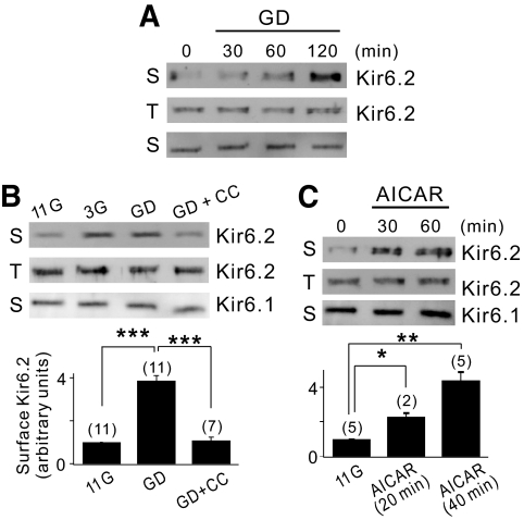
FIG. 3.
Glucose deprivation increases the surface expression level of Kir6.2 through AMPK activation. A: INS-1 cells were incubated with GD medium for the times indicated before surface labeling with a biotin probe. The surface (S) or total (T) expression level of Kir6.2 was assessed by Western blot analysis as described in the online appendix. Kir2.1, a surface channel that does not react on glucose deprivation, was used as a negative control. B: The cells were maintained with 11G, 3G, or GD medium in the presence or absence of 40 μmol/l compound C for 2 h before surface labeling with a biotin probe. The surface expression level of Kir6.2 was quantified by densitometry and expressed as an arbitrary value to that of the cells incubated with GD medium, which was set to 1. Statistical significance was determined by ANOVA. ***P < 0.001. C: The cells were treated with 0.5 mmol/l AICAR-containing 11G medium for the times indicated before surface labeling with a biotin probe. The surface expression level of Kir6.2 was quantified by using the same method in B. Statistical significance was determined by ANOVA. *P < 0.05, **P < 0.01.

