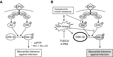
FIG. 7.
Schematic presentation of alteration in EPO-provoked signaling in diabetic myocardium. A: In the normal myocardium (i.e., LETO), EPO phosphorylated both Akt and ERK, but phosphorylation of Akt is sufficient to phosphorylate GSK-3β, resulting in infarct size limitation. B: In diabetic rats (i.e., OLETF rats), Akt phosphorylation by EPO and phosphorylation of GSK-3β by ERK were impaired. Constitutively active GSK-3β is increased, particularly in the mitochondria, which blunts the effect of intracellular signaling leading to GSK-3β phosphorylation.

