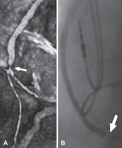
Figure 4).
Bypass graft stenosis (arrows). A Three-dimensional reconstruction of 64-slice multidetector computed tomography image showing bypass graft stenosis at the distal anastamotic site. B Bypass graft stenosis verified by catheter-based coronary angiography. Reproduced with permission from reference 63

