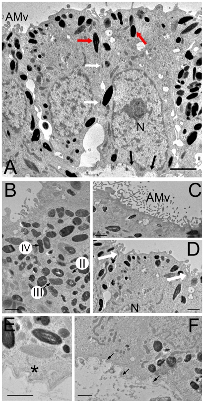
Figure 2. Human iPS-RPE cells are polarized and display classic RPE cell morphology.
(A) Electron micrograph of an iPS-RPE cell monolayer. Human iPS-RPE are pigmented cuboidal epithelial cells with cytoplasmic polarization. Indicated are apical microvilli (AMv), melanin-containing melanosomes (red arrows), the basal nucleus (N), desmosomes (white arrows) and basal lamina (black arrows). (B) Densely packed melanosomes: stages II, III and IV of melanosome maturation are labelled. (C) Microvilli (AMv) extend out from the apical surface of iPS-RPE. (D) Adherens junctions (white arrows) between cells are in the apical portion of the cell, whilst the nucleus (N) is basal. (E) Coated pits (asterisk) are found throughout the plasma membrane. (F) iPS-RPE produce their own basal lamina (indicated by black arrows). Scale bars: A, 2 µm; B-F, 1 µm.

