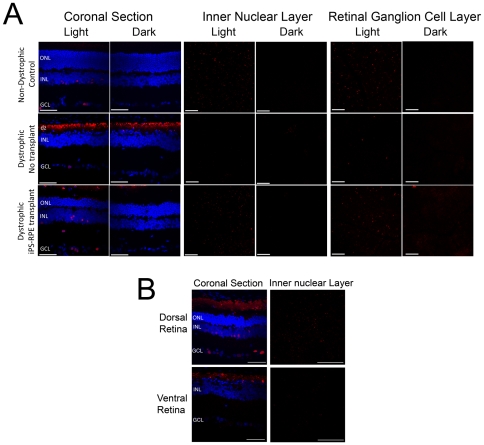
Figure 9. iPS-RPE transplantation preserves the light induced c-Fos response in the RCS dystrophic rat retina.
(A) Age-matched non-dystrophic control, dystrophic no-transplant control and dystrophic rats with iPS-RPE grafts were dark-adapted overnight and sacrificed in the dark or after 90 min exposure to white light (250 µW/cm2). Coronal sections through the retina of dystrophic transplanted, dystrophic non-transplanted and non-dystrophic control RCS rats showing the inner and outer nuclear layers (ONL and INL respectively) and the ganglion cell layer (GCL). Preservation of ONL 13 weeks after iPS-RPE transplant correlates with preservation of the light-induced c-Fos expression (red) in coronal sections of the retina and in representative dorsal whole-mount preparations of the inner nuclear and ganglion cell layers. Note absence of activity in darkness and responsivity to light in the normal and transplanted eyes. c-Fos positive cells in the ganglion cell layer of unoperated RCS eyes match the distribution expected for intrinsically light-responsive melanopsin-containing ganglion cells. DAPI-stained nuclei are shown in blue in the coronal section and the autofluorescent debris zone (dz) is indicated. (B) Light-induced c-Fos activation in the transplanted eye of RCS rats is preferentially preserved in the dorsal retina (the region of the transplant), corresponding with preservation of photoreceptors (ONL) by the iPS-RPE graft. No such preservation is observed in the ventral retina of the transplanted animal. Scale bars: Coronal sections, 50 µm; whole-mount images of the inner nuclear and retinal ganglion cell layers, 200 µm.

