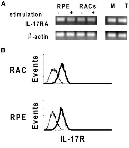
Figure 1.
Expression of IL-17R in RACs and RPE cells. (A) Total RNA was extracted from RACs and RPE cells incubated with or without culture medium containing IFN-γ and TNF-α and also from macrophages (M) or a CD4 T cell line (T). Levels of IL-17RA mRNA were determined by RT-PCR. (B) The receptor expression on RACs and RPE was also tested by flow cytometry at protein level. IL-17R expression by cells is shown by the shift in fluorescence intensity of the specific antibody (thick lines) over the isotype control (thin lines).

