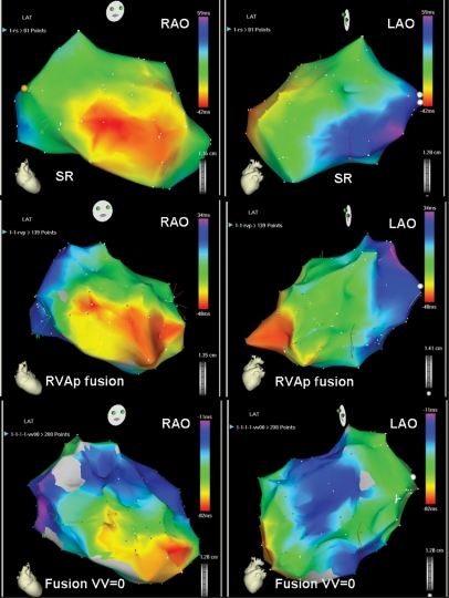
Figure 2.
Example in which echocardiographic AV optimization permitted CRT with fusion. On the top, activation maps during SR showing a mid-septal breakthrough, a postero-lateral most delayed activation area and LVat of 101 ms. On the middle, activation maps during RVA pacing with fusion with the intrinsic depolarization, showing the two septal breakthroughs, a change in the most delayed activation area (antero-lateral) and a shortening in LVat to 82 ms. On the bottom, activation maps with the addition of a third wave front from the CS lead, showing the maintenance of the same septal breakthroughs, a change of the most delayed area of activation towards intermediate positions and a further shortening of LVat to 71 ms.

