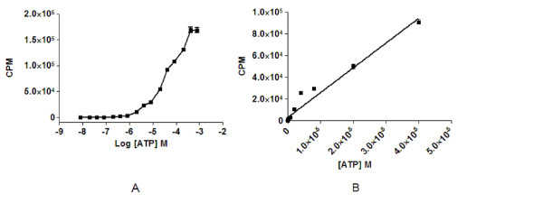
Figure 1.

Detection of exogenous ATP with the luciferase assay. (A) Fifty-five μL of ATP in HMI-9 medium detected by 15 μL of CellTiter-Glo reagent over a dose range resulting in a plateau of the signal at 400 μM. (B) Representation of a linear range of ATP detection with CellTiter-Glo over varying concentrations of ATP. Linearity was approximately 40 μM before the signal plateau. Each concentration was screened in triplicate. The experiment was repeated twice.
