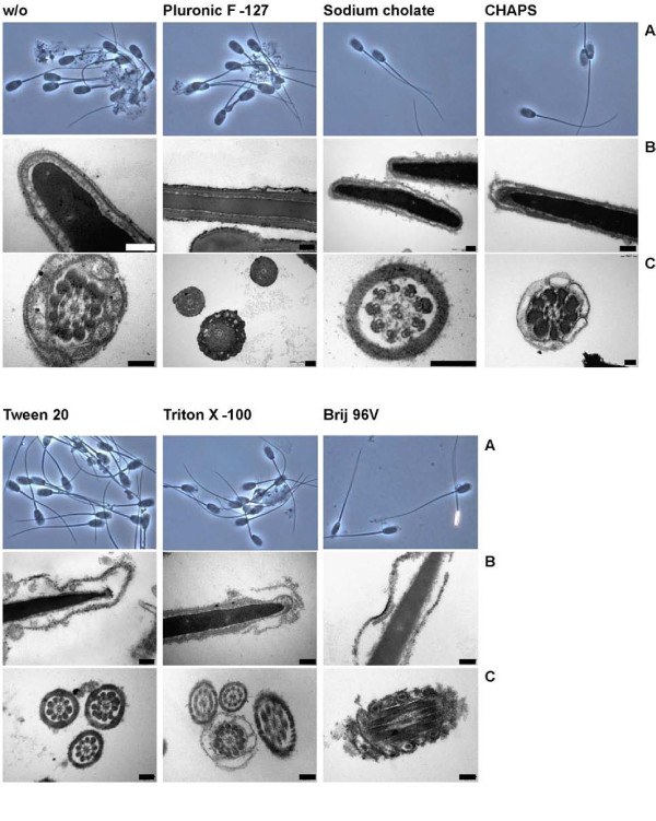
Figure 4.
Morphological patterns of boar sperm membrane lysis after incubation with the indicated detergents for 30 min at 4°C. Sperm samples were observed by light microscopy (A, phase contrast, magnification of 500) as well as by transmission electron microscopy (B, C). For further details see the Materials and Methods section. Each bar represents 200 nm. The quantitative evaluation of the microscopic investigations is given in Tab. 2.

