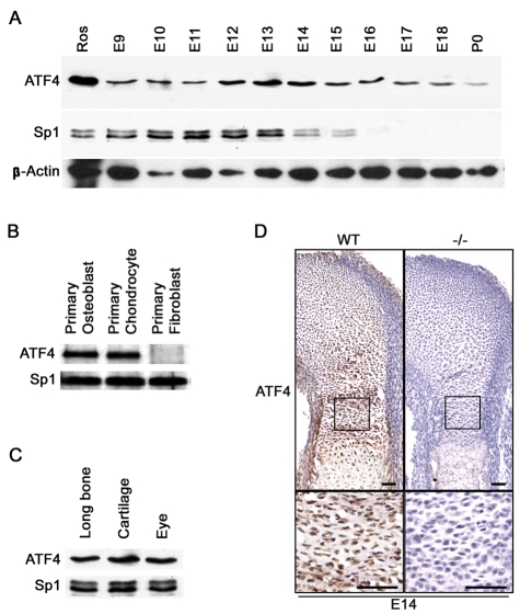
Fig. 1.
Atf4 is expressed in chondrocytes. (A) Western blot of nuclear extracts of mouse embryos (E9-11) and limbs (E12-P0) for Atf4. Sp1 and β-actin were used as a loading control. (B) Western blot of nuclear extracts from the indicated primary cells. (C) Western blot of nuclear extracts from the indicated tissues. (D) Immunohistochemistry of growth plate sections of E14 wild-type (WT) and mutant (Atf4−/−) humeri. The boxed regions are magnified beneath. Scale bars: 50 μm.

