Fig. 4.
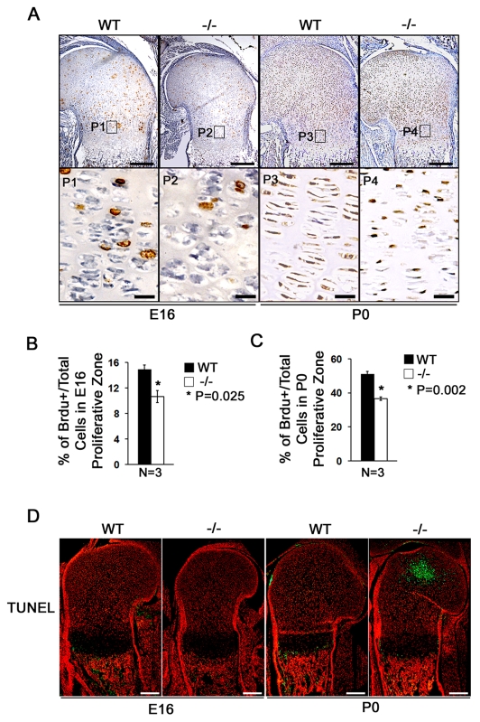
Atf4 is required for chondrocyte proliferation. (A) BrdU immunohistochemistry of E16 and P0 WT and Atf4−/− mouse humerus sections. Boxed regions (P1-P4) are magnified beneath, showing that there are fewer BrdU-positive (brown) proliferative chondrocytes in Atf4−/− growth plates in E16 embryos and P0 pups. (B,C) Quantification of proliferation rate in proliferating chondrocytes, represented by the ratio of BrdU-positive cells normalized to total cells in WT and Atf4−/− E16 (B) and P0 (C) humeri. Error bars indicate s.e.m. (D) TUNEL assay. In E16 WT and Atf4−/− humerus, no apoptotic cells are present in the cartilage, but some TUNEL-positive cells (green) appear in the primary spongiosa. At P0, there are abundant apoptotic cells in the secondary ossification center of the Atf4−/− humerus, but not in the WT. There are also some TUNEL-positive cells in primary spongiosa and hypertrophic chondrocyte zones in both WT and Atf4−/− growth plate. n=5. Scale bars: 0.2 mm in A,D; 0.02 mm in P1-P4 of A.

