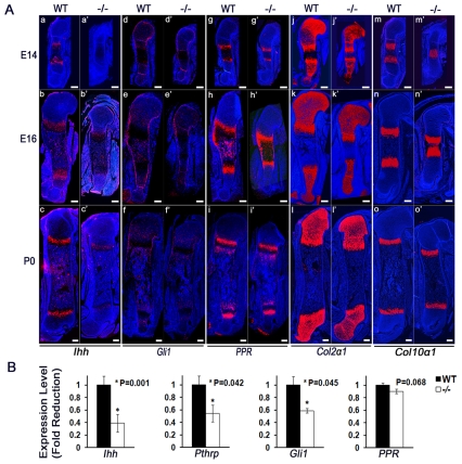
Fig. 5.
Ihh expression is decreased in Atf4−/− cartilage. (Aa-o′) In situ hybridization of sections of E14, E16 and P0 WT and Atf4−/− mouse humeri. Note the decrease in Ihh and Gli1, but normal PPR (Pth1r), expression in Atf4−/− growth plates. Although the Col2a1-positive zones are shorter and the Col10a1-positive zones are slightly longer than their WT counterparts at every stage examined, the expression of Col2a1 and Col10a1 was unchanged in Atf4−/− growth plates. Scale bars: 0.2 mm. (B) qRT-PCR analysis showing decreased levels of Ihh, PTHrP (Pthlh) and Gli1 and normal levels of PPR mRNA in E14 Atf4−/− cartilage. Data are normalized to expression levels in WT cartilage and 18S rRNA (n=3).

