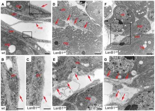
Fig. 5.
Ultrastructural analysis of embryos lacking LanB1. (A-G) Wild-type (A,B) and LanB1def mutant (C-G) embryos at early stage 17. (A) Basal surfaces of the midgut (mg) and hindgut that are closely associated with a layer of visceral musculature (vm). All tissue surfaces exposed to the haemolymph show a uniform layer of BM (arrow in inset). A lamellipodium of a macrophage is also seen that is not accumulating a BM (arrowhead). (B) The CNS is surrounded by a layer of perineural cells that is covered by BM in wild type. (C) In LanB1 mutant embryos only residual ECM material is detected. (D) Gaps between mg and vm cells are apparent in LanB1 mutants (arrows), often filled with unorganised and diffuse ECM material (E, arrows). (F) Section of Malphigian tubule (mt), hindgut with associated vm and a macrophage in LanB1 mutant embryo (F). (G) Close up of boxed region in F. Arrows point to diffuse ECM material at the surface of the mt and vm. Scale bars: 1 μm in F; 2 μm in D; 100 nm in A,C,E,G. hg, hindgut; ma, macrophage; mg, midgut; mt, Malphigian tubule; pn, perineural cell; vm, visceral musculature.

