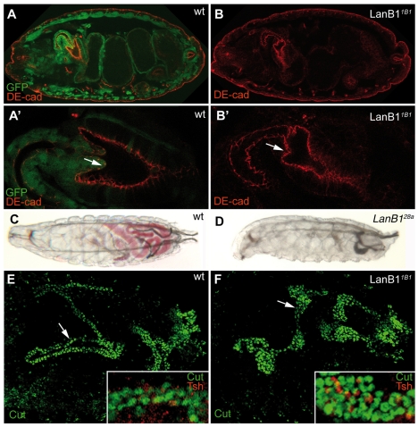
Fig. 7.
Defects in tubulogenesis in LanB1 mutants. (A-B′) Embryos labelled with anti-DE-cadherin (DE-cad, red) and anti-GFP (green) antibodies to visualise the proventricular cells and to mark the balancer chromosome, respectively. (A′,B′) Magnifications of the proventriculus shown in A and B. (A,A′) In wild-type embryos, proventricular cells have moved inward by stage 16 (arrow in A′). (B,B′) This movement fails in homozygous LanB11B1 embryos (arrow in B′). (C,D) Second instar larvae that have been fed with yeast containing red dye. Whereas wild-type larvae can feed normally and show a coloured gut (C), homozygous LanB128a larvae do not (D). (E) Wild-type Malpighian tubules of stage 17 embryos consist of four long tubes, as seen with a principal cell marker, anti-Cut antibody (green). (F) LanB11B1 mutant Malpighian tubules are short and fat, but like the wild type contain principal (green) and stellate (red) cells (E,F, white boxes).

