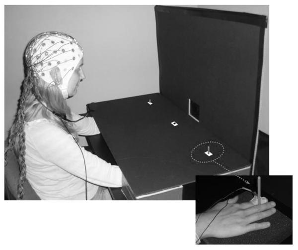
Figure 1.
Stimulus setup used in this study. Sounds were presented from a central loudspeaker that was mounted behind the vertical panel. Red markers were used to indicate the central fixation point and the two lateral saccade target positions. These were located on top of the horizontal panel that was used to cover participants’ hands and forearms. Note that these markers are colored white in this photograph to enhance their visibility. Tactile stimulation was applied to the top phalanx of the left or right index finger. Hands were located underneath the two lateral position markers, and were kept in place by two small vertical sticks, whose top parts were visible on the horizontal panel. The small inset (bottom right) shows the view of the right hand underneath the horizontal panel with the solenoid attached with adhesive medical tape to the index finger and the stick held between index and middle fingers.

