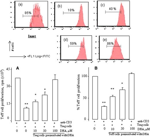
Fig. 1.
DHA reduces Treg cell functions. A: CD4+CD25− T (Teff) cells as responder cells and autologous CD4+CD25+ regulatory T (Treg) cells, both purified from the spleen of mice, were cocultured with soluble anti-CD3/CD28 antibody for 46 h. Treg cells were preincubated or not with increasing concentrations of DHA for 4 h. After incubation, the cells were washed and used for the 46 h proliferation assay. B: The experimental protocol was the same as in A except that Teff cells were preincubated with increasing concentrations of DHA for 4 h. After incubation, the cells were washed and used for the 46 h proliferation assay. In all experiments, cell proliferation was measured by [3H]thymidine incorporation. Results are expressed as mean ± SEM of experiments reproduced at least three times and performed in six replicates from different mice. *P < 0.05 and **P < 0.01: significant difference compared with control cells. Inset shows the Treg suppression assays in which Teff cells were labeled with CFSE (2 µM) and stimulated as described above. In the cell, esterases cleave the acetyl group, leading to the fluorescent diacetylated CFSE. The percentages of residual proliferating cells are indicated in the inset figures. The treatments were as follows: (a), Control Teff cells; (b), Treg cells + Treg cells; (c), Treg cells + Teff cells (preincubated with DHA at 10 μM); (d), Treg cells + Teff cells (preincubated with DHA at 50 μM); (e), Treg cells + Teff cells (preincubated with DHA at 100 μM). Data are representative of three separate experiments.

