Fig. 3.
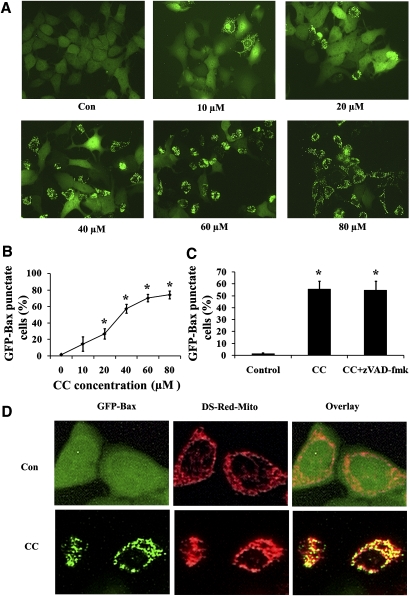
Compound C induced Bax translocation from the cytoplasm to mitochondria in a caspase-independent pathway. A: GFP-Bax stable MCF7 cells were treated with different concentrations of Compound C for 24 h and then visualized by fluorescence microscopy. B: The percentages of GFP-Bax punctate cells were quantitated from four separate visual fields. Values are means ± SEM. * P < 0.05, n = 3. C: GFP-Bax stable MCF7 cells were treated with 40 µM Compound C for 24 h either in the absence or presence of 50 μM zVAD-fmk. The percentages of GFP-Bax punctate cells were visualized by fluorescence microscopy and quantitated from four separate visual fields. Values are means ± SEM. * P < 0.05, n = 3. D: GFP-Bax and Ds-red-mito stable MCF7 cells were treated either with or without 40 µM Compound C for 24 h. The cells were then visualized by fluorescence microscopy.

