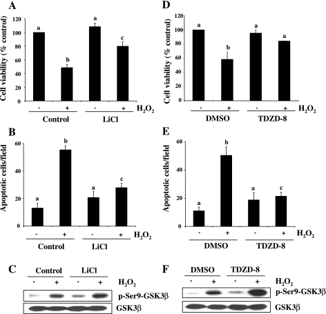
Fig. 9.
Effect of GSK-3β inactivation on RPTC survival following oxidant injury. Serum-starved RPTC were pretreated with LiCl (20 mM) and TDZD-8 (40 μM) for 1 h and then exposed to 1 mM H2O2 for 4 h (A, B, D, and E) or for 30 min (C and F). Cell viability and apoptosis were assessed by the MTT assay (A and D) and DAPI staining (B and E), respectively. Values are means ± SD of 3 independent experiments conducted in triplicates and expressed as the percentage of control (A and D) or total apoptotic cells per field (B and E). Bars with different letters are significantly different from one another (P < 0.05). Cell lysates were prepared for immunoblot analysis using antibodies against p-Ser9-GSK-3β and GSK-3β (C and F). Representative immunoblots from 3 experiments are shown.

