Fig. 5.
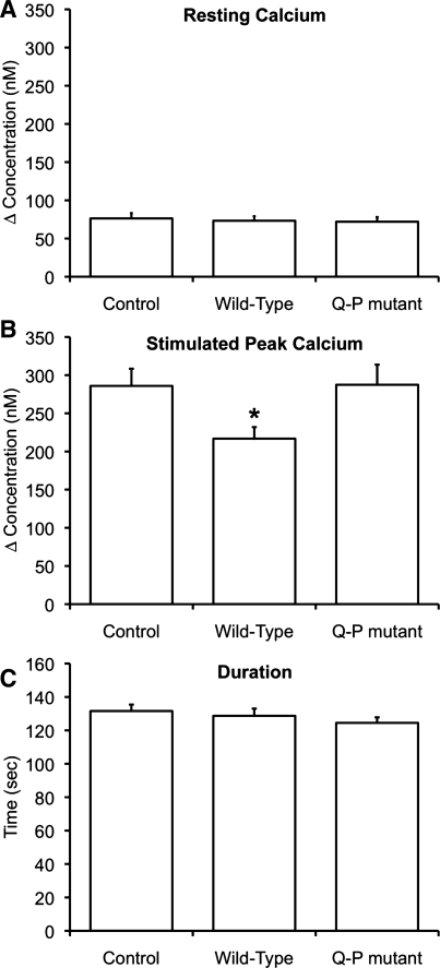
Expression of PC1-C, but not PC1-C-Q4215P, alters intracellular Ca2+ signaling. MDCK cells were loaded with fura 2-AM, and changes in intracellular Ca2+ levels were monitored upon addition of extracellular ATP in the absence of extracellular Ca2+. In all cases, the values plotted represent the average of the response measured in 92-254 cells from experiments performed on at least 5 different days. Control, experiments where cells were mock transfected with empty vector. A: resting Ca2+ levels were unchanged by the expression of PC1-4183-4270 (wild-type) or PC1-4183-4270-Q4215P (Q-P mutant). B: peak Ca2+ release was attenuated after addition of 3 μM ATP in cells expressing PC1-C wild-type, but not the Q-P mutant, presumably because expression of PC1-C disrupts the normal interaction between full-length PC1 and PC2 in MDCK cells. C: duration of the response to 3 μM ATP was similar in cells expressing PC1-C wild-type and the Q-P mutant. *Statistically significant (P <0.05).

