Fig. 1.
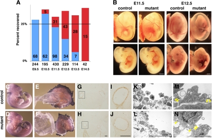
Neural crest-specific disruption of Bmpr1a leads to acute death by embryonic day (E) 12.5 without overt abnormality in vessel formation. A: recovery of mutants with no overt abnormalities (blue) vs. those that have died or exhibiting hemorrhage (red) are plotted as a function of gestation age. Actual number of embryos recovered is given in each bar. Total number of embryos is given above gestation age. B: external view of embryos at E11.5 (left 4 panels) and E12.5 (right 4 panels). Top, embryos before removal of yolk sac; bottom, dissected embryos from respective yolk sac. C–F: platelet endothelial cell adhesion molecule staining at E11.5. G–J: α-smooth muscle actin staining at E11.5. G and H: third pharyngeal arch (PhA) artery in control and mutant, respectively. I and J: higher magnification of G and H, respectively. Bar = 100 μm. K–N: transmission electron-microscopy images of vessels. K and L: third PhA artery of control and mutant embryo, respectively. M and N: higher magnification of rectangle areas in K and L, respectively; p, pericyte; e, endothelial cells; yellow arrowhead, junctions of endothelial cells. Bar = 1 mm for B, 20 μm for K and L, and 2 μm for M and N.

