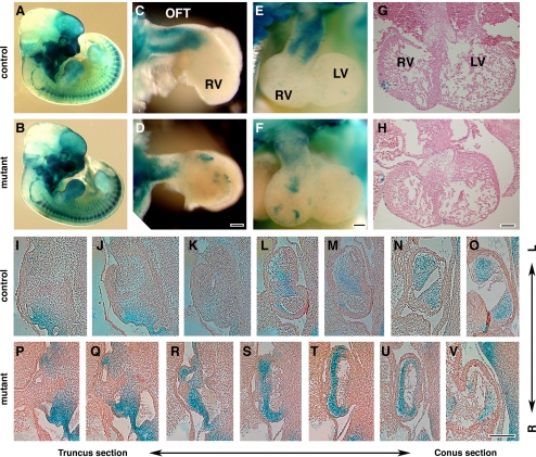
Fig. 2.
Cardiac neural crest cells migrate into the outflow tract (OFT) with altered distribution in mutant. A and B: visualization of neural crest cells derivatives at E11.5 by X-gal staining (blue) to visualize ROSA26-derived β-gal expression. C–F: whole-mount view of the X-gal stained heart after removal of atrium. C and D: lateral view. E and F: anterior view. G and H: transverse sections of E11.5 hearts stained with hematoxylin-eosin. I–V: frontal sections of OFTs from X-gal-stained embryos counterstained with eosin. LV, left ventricle; RV, right ventricle. Bar = 100 μm.

