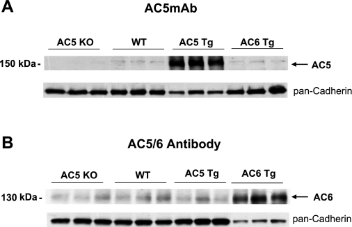
Fig. 2.
Western blot analysis of AC5MAb with mouse heart. A: AC5 protein is detected by immunoblotting and is absent in AC5 KO and increased in AC5 TG hearts. The level of AC5 was similar to WT in the AC6 TG mouse heart. The amount of protein loaded from AC5 TG heart is 50% of the amount from AC5 KO, WT, and AC6 TG hearts. B: immunoblot using an AC5/6 commercial antibody. The AC levels were not different in hearts of AC5 KO, WT, and AC5 TG but increased in AC6 TG hearts, indicating that this antibody may primarily detect only AC6 protein. The amount of protein loaded from AC6 TG heart is 30% of the amount from AC5 KO, WT, and AC5 TG hearts. The loading control is shown using pan-cadherin antibody.

