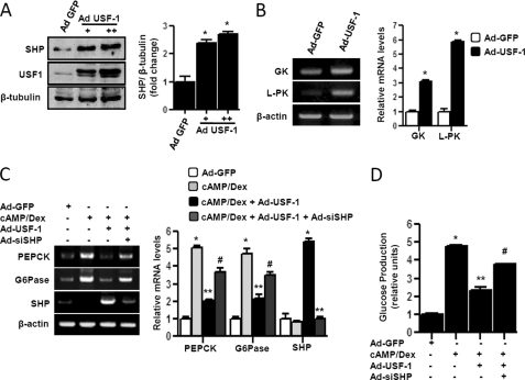
FIGURE 5.
USF-1 represses gluconeogenesis via induction of SHP gene expression in primary hepatocytes. A and B, primary rat hepatocytes were infected with adenovirus (Ad) GFP (50 m.o.i.) or Ad-USF-1 (+ = 25 m.o.i., ++ = 50 m.o.i.) for 36 h. Cells were harvested for Western blot analysis using indicated antibodies (A), or total RNA was isolated for semi-quantitative RT-PCR analysis (B). Data represent mean ± S.D. of three individual experiments. *, p < 0.001 compared with Ad-GFP-treated cells. C, cells were infected with Ad-GFP, Ad-USF-1, or Ad-siSHP followed by Ad-USF-1 for 36–48 h preceding cAMP (500 μm) and Dex (100 nm) treatment for 3 h. Total RNA was isolated for semiquantitative RT-PCR analysis. Data represent mean ± S.D. of three individual experiments. *, p < 0.001, **, p < 0.05, and #, p < 0.001 compared with Ad-GFP-infected cells, cAMP/Dex treatment, and Ad-USF-1 infected cells, respectively. D, measurement of glucose production. Experiments were performed as described in C, using glucose-free media supplemented with gluconeogenic substrate sodium lactate (20 mm) and sodium pyruvate (1 mm). Data represent mean ± S.D. of four individual experiments. *, p < 0.001; **, p < 0.001, and #, p < 0.001 compared with Ad-GFP infected cells, cAMP/Dex treatment, and Ad-USF-1 infected cells, respectively. G6Pase, Glc-6-Pase.

