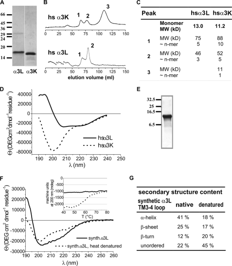
FIGURE 4.
Secondary structure and association of recombinant and synthesized TM3–4 loops. A, SDS-polyacrylamide gel electrophoresis and Coomassie stain to verify integrity and purity of expressed TM3–4 loops. The subunits and size markers are indicated. B, size exclusion chromatography of recombinant TM3–4 loops on a Sephacryl S-200 column. Peaks indicating different oligomerization states are labeled. C, summary of size exclusion chromatography data. D, CD spectra of TM3–4 loops of recombinant α3L and α3K from expression in E. coli. E, SDS-polyacrylamide gel electrophoresis and Coomassie stain to verify integrity and purity of the synthesized α3L TM3–4 loop. The size markers are indicated. F, CD spectra of the synthetic α3L TM3–4 loop before and after thermal denaturation. Inset, thermal unfolding and cooling curves of synthetic α3L TM3–4 loop. Ellipticity at 200 nm is plotted versus temperature. Note melting point of highly ordered structure at ∼58 °C. Dashed line, heating, solid line, cooling. G, summary of secondary structure analysis data of synthetic α3L TM3–4 loop. MW, molecular weight.

