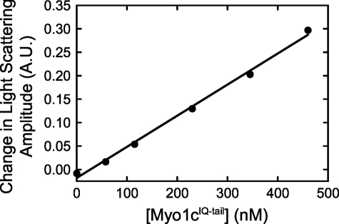
FIGURE 1.
Amplitude change of light scattering of LUVs increases linearly with myo1cIQ-tail concentrations. LUVs containing 2% PtdIns(4,5)P2 (115 μm total lipid) were pre-equilibrated with increasing concentrations of myo1cIQ-tail and mixed with either buffer alone or 58 μm InsP6. Blank-subtracted transients were fit to a single exponential function, and the amplitudes of the fits are plotted as a function of the myo1cIQ-tail concentration. The solid line represents a linear fit to the data. Concentrations are after mixing.

