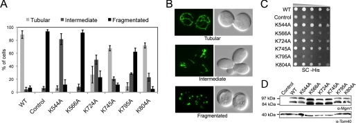
FIGURE 4.
s-Mgm1 mutants defective in IM binding and stimulated GTPase activity have impaired function in vivo. A, the mitochondrial morphology of complementation strains (Control represents no Mgm1 expressed) was scored by fluorescence microscopy of a mitochondrially targeted green fluorescent protein. 300 cells were counted for each mutant complementation experiment. B, shown are representative micrographs of mitochondrial green fluorescent protein morphology that categorizes tubular (upper panel), intermediate (middle panel), and fragmented (lower panel) morphology. C, serial dilution onto synthetic complete medium (SC) demonstrated growth rates of lipid-binding mutants (Control represents no Mgm1 expressed). D, Western blot analysis of mutant strains (Control represents no Mgm1 expressed) demonstrated that Mgm1 was processed in all mutants tested (upper panel) and expressed to similar levels normalized to Tom40 levels (lower panel).

