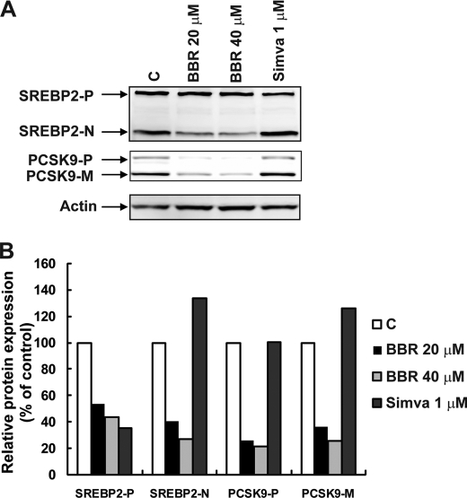
FIGURE 7.
Reduction of SREBP2 protein by BBR treatment is partially responsible for reduced PCSK9 expression. A, HepG2 cells were treated with BBR for 24 h at concentrations of 20 and 40 μm. Total cell lysate was isolated for Western blot analysis of SREBP2 and PCSK9. B, intensities of the bands of precursor SREBP2 (SREBP2-P), nuclear SREBP2 (SREBP2-N), precursor PCSK9 (PCSK9-P), and mature PCSK9 (PCSK9-M) were quantified using an imaging program of Kodak Imaging Station 4000R. Values were normalized to β-actin and were graphed relative to untreated cells. The figure shown is representative of three separate experiments with similar results.

