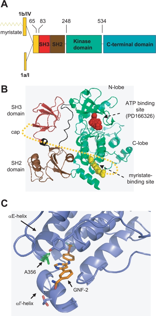
FIGURE 1.
A, domain structure of Abl family members (5). The numbers indicate amino acid residues in c-Abl 1b, and the recombinant protein constructs used in this study encompass amino acids 65–534, 83–534, and 248–531. B, ribbon representation of the c-Abl kinase NH2-terminal half residues, including the SH3, SH2, and kinase domains (Protein Data Bank code 1OPK) (7). The NH2-terminal cap (amino acids 2–79) is indicated by dotted lines (8). The myristate-binding site and ATP binding pocket are indicated by arrows. C, ribbon representation of an enlarged view of GNF-2 (colored gold) bound to the c-Abl myristate binding site. The location of Ala356 is indicated.

