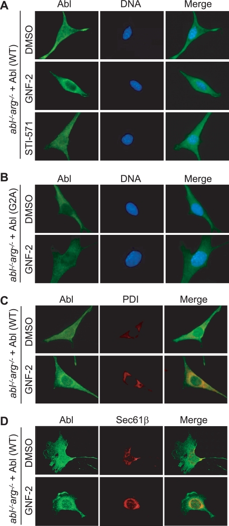
FIGURE 6.
GNF-2 induces translocation of the myristoylated c-Abl to the ER. A and B, the indicated cells were plated on coverslips and grown in DMEM containing 10% fetal bovine serum. The cells were treated with either DMSO or 10 μm compound for 1 h, and c-Abl proteins were visualized by indirect immunofluorescence using an anti-c-Abl antibody (8E9) and Alexa Fluor 488 goat anti-mouse IgG. 4′,6-Diamidino-2-phenylindole was used to stain the nucleus. C, wide field fluorescence images. The cells were double-stained for c-Abl (green) and protein-disulfide isomerase (red) following treatment with either DMSO or 10 μm GNF-2 for 1 h. D, confocal fluorescence images. The cells were double-stained for c-Abl (green) and Sec61β (red) following treatment with either DMSO or 10 μm GNF-2 for 1 h. It is evident that the intense green fluorescence around the nucleus in GNF-2-treated cells colocalizes with the red fluorescence.

