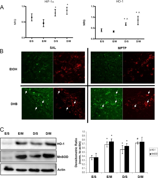
FIGURE 2.
HIF-1α and its downstream target HO-1 are up-regulated by DHB in the SN; up-regulation is maintained in the presence of MPTP. A, RT-PCR analysis of Hif-1α and its downstream target Ho-1 were performed on midbrain SN tissue obtained 6 h post-treatment; means ± S.D. are shown. EtOH/SAL (E/S), EtOH/MPTP (E/M), DHB/SAL (D/S), and DHB/MPTP (D/M). *, p < 0.05 significantly different from EtOH/MPTP; ⋀, p < 0.05 significantly different from EtOH/SAL (n = 4). B, 7-μm paraffin-embedded sections of SN, obtained from mice harvested at 24 h post-treatment, were stained for HIF-1α (green) and TH (red); white arrows point to HIF-1α localized within representative DAergic SN neurons. C, 100 μg of total protein extracts prepared from midbrain SN homogenates were loaded onto 4–12% BisTris gels and electrophoresed prior to transfer onto polyvinylidene difluoride membranes and subsequently immunoblotted for HO-1 and MnSOD; actin was used as a loading control, a representative blot is shown in the left panel and quantification of at least 3 separate immunoblots (means ± S.E.) is shown in right panel. *, p < 0.05 significantly different from EtOH/SAL; #, p < 0.05 significantly different from DHB/SAL.

