FIGURE 7.
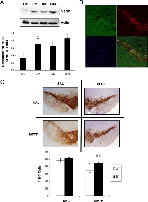
VEGF overexpression protects against MPTP-induced nigral cell death. A, 100 μg of total protein extracts prepared from midbrain SN homogenates were loaded onto 4–12% BisTris gels and electrophoresed prior to transfer onto polyvinylidene difluoride membranes and subsequent immunoblotting for VEGF; actin was used as a loading control, a representative blot is shown in upper panel and quantification of at least 3 separate immunoblots (means ± S.E.) is shown in the lower panel. *, p < 0.05 significantly different from EtOH/SAL; #, p < 0.05 significantly different from DHB/SAL. B, 20-μm sections of SN obtained from VEGF transgenic mice were stained for VEGF (green) and TH (red); overlay of VEGF and TH staining demonstrates co-localization of VEGF in DAergic neurons (yellow-orange). C, brain scans show TH-stained DAergic neurons of saline (SAL)-treated mice from the wild-type and VEGF transgenic groups (upper scans). Bottom scans show the mice from the corresponding groups treated with MPTP and then allowed to recover for 7 days before sacrifice. Quantification of TH-positive cell counts is depicted in the graph. *, p < 0.05 between SAL/SAL and SAL/MPTP; &, p < 0.05 between VEGF WT/MPTP and VEGF TG/MPTP; #, p < 0.05 between VEGF TG/SAL and VEGF TG/MPTP (mean ± S.E.; n = 4).

