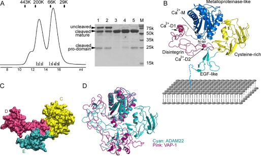
FIGURE 1.
Structure of the ADAM22 ectodomain. A, gel filtration profile in a calibrated Superdex 200 column showing that secreted ADAM22 ectodomain exists as a mixture of three forms: the uncleaved propeptide-containing ADAM22, the non-covalent complex between propeptide and mature ADAM22, and the mature ADAM22 without propeptide. The eluted peaks were analyzed by SDS-PAGE. Lane M, molecular size. B, ribbons diagram showing the topology of the ADAM22 ectodomain. The M, D, C, and E domains are shown in blue, magenta, yellow, and cyan, respectively. The three putative calcium ions are colored the same as their host domains. EGF, epidermal growth factor. C, surface diagram of the D, C, and E domains showing that these three domains have seamless interfaces and are an integral module. D, superimposition of ADAM22 and VAP-1 based on overlapping their M domains showing a significant difference in spatial relation between domain M and the rest of the molecule.

