FIGURE 5.
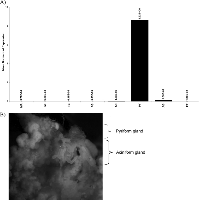
Pyriform glands are fingerlike structures that express PySp1 mRNA at high levels. A, real time quantitative PCR analysis was used to determine the mRNA expression pattern of the novel fibroin in a variety of different tissues. Total RNA was isolated from the major ampullate gland (MA), minor ampullate gland (MI), tubuliform (TB), flagelliform (FL), aciniform (AC), pyriform (PY), aggregate (AG), and fat tissues (FT). Equivalent amounts of total RNA were reversed transcribed using Superscript III and aliquots used for quantitative reverse transcriptase-PCR. Reactions were performed in triplicate and normalized internally using the black widow actin mRNA. Data are representative of experimental results obtained from two independent trials. B, pyriform glands were dissected from black widow spiders (upper right, pyriform gland removed from a single spider and photographed on a Leica MZ16 dissecting microscope at ×10 magnification; lower left, aciniform tissue is also shown in the field of view; fat tissue is surrounding both glands).

