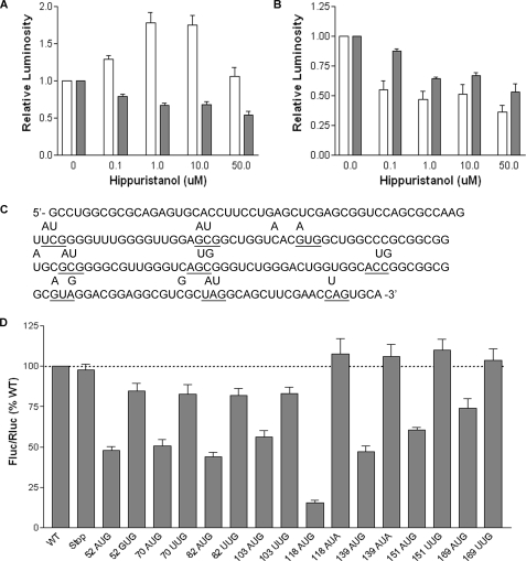
FIGURE 7.
Ribosome scanning occurs between nucleotides 103 and 118 of the SHMT1 5′-UTR. MCF-7 cells were transiently transfected with polyadenylylated bicistronic mRNAs containing the hepatitis C virus IRES (A) or the SHMT1 5′-UTR (B) and then treated with the indicated amount of hippuristanol. Following 11 h of treatment, Fluc (white bars) and Rluc (dark bars) expression was quantified as described under ”Experimental Procedures.“ The relative luminosity in untreated cells was given a value of 1.0. The data represent the average of three independent experiments ± S.E. C, the sequence of the SHMT1 5′-UTR indicating the positions of the inserted open reading frames. The location of each start and stop codon is underlined, and the letters above the underlined nucleotides indicate changes to the wild-type sequence that were made by site-directed mutagenesis. D, MCF-7 cells were transiently transfected with bicistronic mRNAs containing the mutated SHMT1 5′-UTRs. The number of each mutant represents the position of the A in the AUG or AUA, or the U in the UUG. The relative ratio of Fluc/Rluc for each the wild-type (WT) bicistronic mRNA was given a value of 100%. The data represent the average of at least three independent experiments ± S.E.

