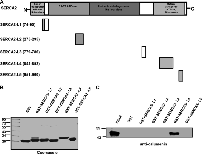
FIGURE 9.
Region of SERCA2 interacting with calumenin. A, schematic representation of mouse SERCA2 and its five luminal loop region constructs. The loops are marked by their amino acid positions, L1–L5 corresponding to mouse SERCA2 domains that face the luminal side of SR. B, GST and GST-SERCA2 fusion proteins L1–L5 (predicted approximate molecular sizes ∼28.0, ∼28.3, ∼26.8, ∼30.6, and ∼27.0 kDa, respectively) were analyzed by SDS-PAGE and stained with Coomassie Blue. C, pulldown assay was performed using equivalent amounts of control GST protein and GST-SERCA2 fusion peptides L1–L5 bound to Sepharose 4B by incubating with cardiac homogenates. The pulldown samples were separated by SDS-PAGE and immunoblotted with anti-calumenin antibody.

