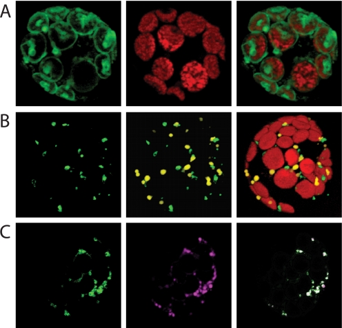
FIGURE 6.
Localization of AtNDT1- and AtNDT2-GFP proteins in tobacco leaf protoplasts. Tobacco protoplasts were prepared as given under “Experimental Procedures” and transformed using polyethylene glycol. Fluorescence was visualized after 18 h of incubation by fluorescence microscopy. Chloroplasts were visualized by their autofluorescence. A, shown is localization of AtNDT1-GFP. B, shown are localization of AtNDT2-GFP and cotransformation with SKL-DSred. C, shown is the localization of AtNDT2-GFP and coincubation with the Mito Tracker dye.

