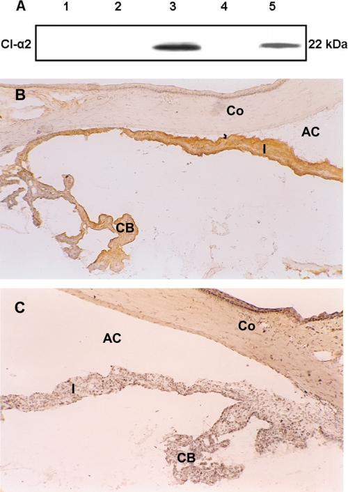
FIGURE 4.
Localization of MAA [CI-α2 (22 kDa)) in the eye. A, Western blot analysis using polyclonal antibody against bovine MAA (CI-α2 (22 kDa)) to detect MAA in different rat ocular tissues. Samples were analyzed on 14% SDS-PAGE. Total protein (40 μg) from cornea (lane 1), retina (lane 2), iris and CB (lane 3), and RPE and choroid (lane 4) were used. Purified bovine MAA (CI-α2 (22 kDa)) loaded (5 μg) in lane 5 was used as the positive control. B and C, MAA (CI-α2 (22 kDa)) in the rat eye (frozen sections). Naive rat eye was examined immunohistochemically for MAA using polyclonal antibody against bovine MAA. B, MAA is constitutively expressed on the iris (I) and ciliary body (CB); MAA was not detected on the cornea (Co). No staining was observed in the control section stained with normal rabbit serum (C). The data shown are representative of three separate experiments. AC, anterior chamber. Objective magnification was ×10.

