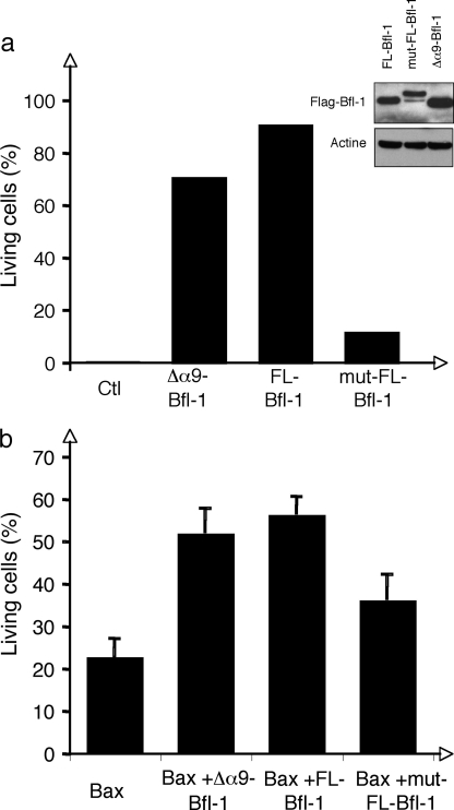
FIGURE 6.
Role of the amphipathic helix α9 for Bfl-1 anti-apoptotic activity. a, NIH3T3 cells were stably transduced with empty vector (Ctl), FLAG-Δα9-Bfl-1pEGZ, FLAG-FL-Bfl-1pMIG, or FLAG-mut-FL-Bfl-1pMIG. Apoptosis was induced by serum deprivation. Cell death was measured by propidium iodide staining and FACS analysis 72 h following serum withdrawal. Data are representative of three independent experiments. Expression of FL-, mut-, and Δα9-Bfl-1 proteins was controlled by Western blot using anti-FLAG antibody and is shown in the inset. b, HeLa cells were transiently transfected with Bax-pEGFP-N1 vector (Bax), Bax-pEGFP-N1 + FLAG-Δα9-Bfl-1pEGZ (Δα9-Bfl-1), Bax-pEGFP-N1 + FLAG-FL-Bfl-1pEGZ (FL-Bfl-1) or Bax-pEGFP-N1 + FLAG-mut-FL-Bfl-1pEGZ (mut-FL-Bfl-1). Cell death was measured by propidium iodide staining at 48 h. Data are presented as percent of living cells among GFP-positive cells. Data are presented as a mean of five (Bax, FL-Bfl-1, and mut-FL-Bfl-1) or two (Δα9-Bfl-1) independent experiments.

