FIGURE 1.
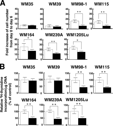
Effects of TPA on the in vitro growth and DNA synthesis of melanoma cell lines. A, seven melanoma cell lines (WM35, WM39, WM98-1, WM115, WM164, WM239A, WM1205Lu) were plated at a density of 1 × 10⁵ cells in 10-cm tissue culture dishes, and the following day (day 0) the cells were treated with either 0.1% DMSO (control; open bars) or 100 nm TPA (filled bars). On day 6 the triplicate dishes were trypsinized, and the cell number was counted. Data are expressed as fold increase of cell number from day 0 to day 6 and are shown as mean ± S.D. (n = 3; *, p < 0.05; **, p < 0.01). B, the seven melanoma cell lines used in A were seeded at 104 cells/well in 24-well plates and grown for 24 h in EMEM containing 5% FCS. Cells were then treated with either 0.1% DMSO (control; open bars) or 100 nm TPA (filled bars) and cultured for another 24 h. [3H]Thymidine was added for the last 6 h of the incubation, and [3H]thymidine incorporation was determined by scintillation counting. [3H]Thymidine incorporation/mg DNA was calculated. Data are expressed as percentage of control and are shown as mean ± S.D. (n = 3; **, p < 0.01). The results shown are representative of three independent experiments.

