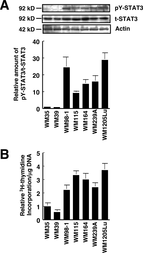
FIGURE 2.
STAT3 is activated in human melanoma cells. A, whole cell extracts from the seven human melanoma cell lines used in Fig. 1 were prepared, and the phosphorylation level of pY-STAT3 as well as the expression of total STAT3 (t-STAT3) were determined by immunoblot analysis using anti-pY-STAT3 and anti-STAT3 antibodies, respectively. Expression of actin was also examined (top three panels). The relative amount of pY-STAT3/t-STAT3 was calculated (bottom panel). The ratio of pY-STAT3 to total STAT3 in WM35 cells is represented as 1. B, the seven melanoma cell lines used in A were seeded at 104 cells/well in 24-well plates and grown for 48 h in EMEM containing 5% FCS. [3H]Thymidine was added for the last 6 h of the incubation, and [3H]thymidine incorporation was determined by scintillation counting. [3H]Thymidine incorporation/mg DNA was calculated for each cell line, and data are expressed as relative [3H]thymidine incorporation/mg of DNA. The value of WM35 cells is represented as 1, and data are shown as mean ± S.D. The results shown are representative of three independent experiments.

