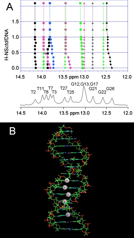
FIGURE 6.
Sites of target DNA involved in H-NSctd recognition. A, 400-MHz NMR spectrum of the imino resonances of the 15-bp DNA alone and in the presence of increasing amounts of H-NSctd. The DNA numbering schemes are as follows: 1CTTACATTCCTGGCT15, 5′ to 3′; and 30GAATGTAAGGACCGA16, 3′ to 5′. B, model of the 15-bp DNA fragment. The spheres represent the imino protons that show the largest chemical shift differences upon addition of H-NSctd.

