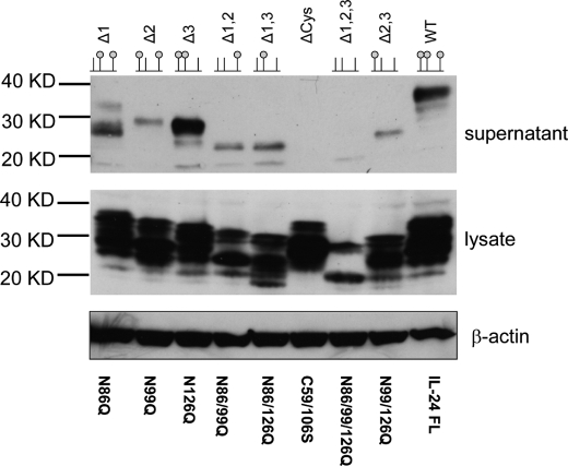
FIGURE 3.
Western blot of SDS gel showing soluble secreted human IL-24 (supernatant) and insoluble, intracellular IL-24 (lysate). The top row shows the identity of the mutation and a schematic lollipop representation of the mutation. The small green balls correspond to the occupancy of a glycan moiety. In almost every case, the lysate shows immune-reactive protein at various stages of processing. β-Actin was used as a positive control for sample loading.

