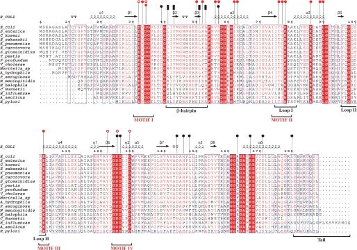
FIGURE 2.
Multiple sequence alignment of KdcC homologs. The secondary structure of E. coli KdsC is shown schematically above the alignment. Residues in the β2–β3-hairpin and outside of the hairpin that form the tetramerization interface are designated by the black bars and circles, respectively. Residues that are involved in coordination of Mg2+ and phosphate are shown by the open red circles and those that are involved in Kdo recognition are shown by the filled red circles. Four conserved HADSF motifs are designated. β-turns are designated as TT.

