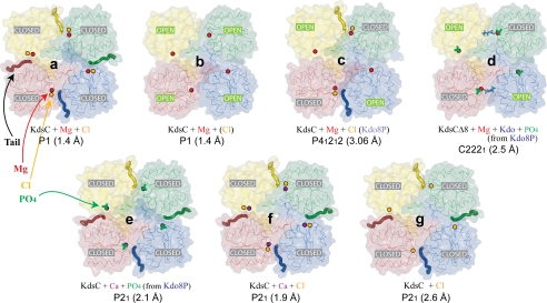
FIGURE 3.
A representative view of all seven tetrameric KdsC structures. The conformation of the active site (open or closed) for each monomer is specified. The positions of Mg2+, Ca2+, and Cl− ions are shown as the red, purple, and orange spheres, respectively. Kdo8P (blue), Kdo (blue), and the phosphate (green) are shown as stick models. Captions designate the contents of each crystal forms, their space groups, and resolutions (for more details, see Tables 1 and 2). The compounds present in the crystallization mixture but not observed in the protein structure are written in parentheses.

