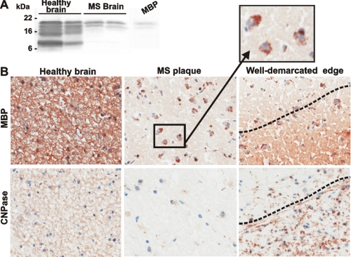
FIGURE 1.
Representative immunostaining of the healthy and MS brain sections. A, protein extracts (25 μg of total protein each) from a healthy total brain and medulla oblongata (left and right lines, respectively) and from the plaque area of the two MS brain samples were analyzed by Western blotting with the MBP antibody. The extracts were purchased from BioChain Institute (Hayward, CA). The representative samples are shown. Right lane, purified MBP (10 ng). B, immunostaining of the brain sections with the MBP and 2′,3′-cyclic nucleotide 3′-phosphodiesterase (CNPase) antibodies. The staining with the primary antibodies was followed by a diaminobenzidine-based detection method. A well demarcated edge (right) is visible between the plaque and the normal brain area. Magnification, ×160. Inset, the phagocytic macrophages (identified by their characteristic size and morphology) co-localize with fragmented MBP (brown) in the plaque. The identity of the macrophages was additionally confirmed using an antibody against the pan macrophage F4/80 marker (Pharmingen).

