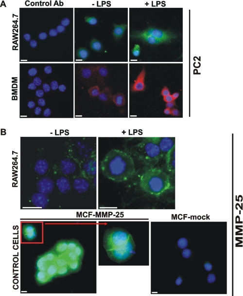
FIGURE 5.
Immunostaining of PC2 and MMP-25. A, macrophage RAW264.7 cells and the murine BMDM were grown in the LabTek chamber slides. The cells were left intact (−LPS) or stimulated 24 h with LPS (1 μg/ml). The cells were fixed, permeabilized, and stained with the PC2 antibody (Ab) followed by the secondary antibody conjugated with Alexa Fluor 488 (green) or Alexa Fluor 594 (red). Staining with the preimmune rabbit serum (control antibody) was negative. The nuclei were stained with 4′,6-diamidino-2-phenylindole (blue). B, macrophage RAW264.7 cells were grown in the LabTek chamber slides. The cells were left intact (−LPS) or stimulated 24 h with LPS (1 μg/ml). The cells were fixed and stained with the MMP-25 antibody. Breast carcinoma MCF7 cells stably transfected with MMP-25 (MCF-MMP-25) or the empty plasmid (MCF-mock) were used as controls. Inset, cell surface staining of MMP-25. The nuclei were stained with 4′,6-diamidino-2-phenylindole (blue). Original magnification ×400; the bar, 10 μm.

