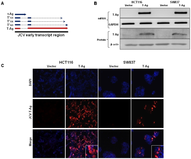
Figure 1. JCV T-Ag expression in transfected colorectal cancer cells.
(A) An illustration of the JCV early transcript region that codes for 5 early transforming proteins, T-Ag, t-Ag and the 3 splice variants T'165, T'136, T'135. T-Ag is predominant protein expressed after transfection (marked in red). (B) RT-PCR and Western immunoblotting gel images depicting JCV T-Ag specific mRNA and protein expression in stably transfected cells (indicated as T-Ag), while no T-Ag expression was observed in control cell lines (V, vector transfected). GAPDH (RT-PCR) and β-actin (WB) were used as loading controls. (C) Immunofluorescence staining with JCV T-Ag antibody shows nuclear expression of JCV T-Ag in transfected HCT116 and SW837 cells lines. The images were taken at a final magnification of 630×.

