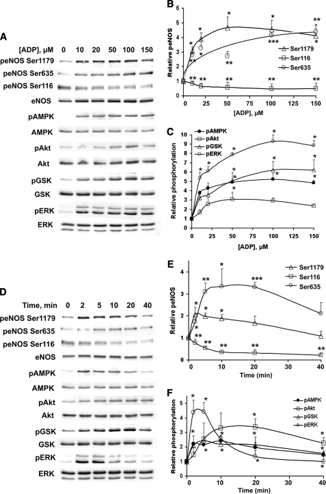
FIGURE 1.
Dose response and time course for ADP-mediated phosphorylation responses in endothelial cells. Lysates prepared from ADP-treated BAEC were resolved by SDS-PAGE and analyzed in immunoblots probed with antibodies directed against phospho-eNOS Ser1179, phospho-eNOS Ser635, phospho-eNOS Ser116, phospho-AMPK, phospho-Akt, phospho-GSK3β, and phospho-ERK1/2. Equal loading was confirmed by immunoblotting with antibodies directed against eNOS, AMPK, Akt, GSK3β, and ERK1/2. Shown on the left are representative immunoblots, and on the right are quantitative plots derived from pooled data. Each point in the graphs represents the mean ± S.E. of four independent experiments that yielded similar results. * indicates p < 0.05. ** indicates p < 0.01. A, BAEC were treated with the indicated concentrations of ADP for 5 min. B, quantitative analysis of phospho-eNOS as a function of concentration. C, quantitation of phospho-AMPK, phospho-Akt, phospho-GSK3β, and phospho-ERK1/2 as a function of ADP concentration. D, BAEC were treated with 15 μm ADP for the indicated times. E, quantitation of phospho-eNOS as a function of treatment time. F, quantitative analysis of phospho-AMPK, phospho-Akt, phospho-GSK3β, and phospho-ERK1/2 abundance as a function of treatment time. p, phospho.

