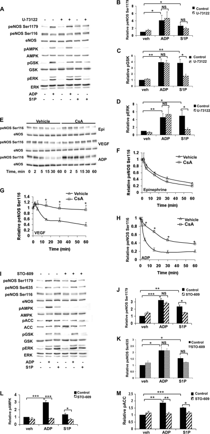
FIGURE 4.
Endothelial responses to ADP and S1P after inhibition of phospholipase C, calcineurin, or CaMKKβ. A, BAEC were treated with the PLC inhibitor U-73122 (10 μm) for 30 min and then stimulated for 5 min with ADP (50 μm) or S1P (100 nm). Immunoblots were probed with antibodies as indicated. Equal loading was confirmed using antibodies directed against total eNOS, AMPK, GSK3β, and ERK1/2. Shown is a representative immunoblot from four individual experiments that yielded similar results. Quantitative analysis of pooled data is shown for the relative phosphorylation of eNOS Ser1179 (B), GSK3β (C), and ERK1/2 (D). E, cell lysates were prepared from BAEC pretreated for 30 min with cyclosporine (100 nm) and then stimulated for the times indicated with epinephrine (1 μm), VEGF (20 ng/ml), or ADP (50 μm). Lysates from BAEC were resolved by SDS-PAGE and probed using antibodies directed against phospho-eNOS Ser116 and eNOS. Shown here is an immunoblot representing three individual experiments with equivalent results. Analysis of pooled data quantitating relative eNOS Ser116 phosphorylation after treatment with epinephrine, VEGF, or ADP is shown in F–H, respectively. * indicates p < 0.05 compared with agonist-mediated levels of eNOS Ser116 phosphorylation in the absence of cyclosporine. I, BAEC were incubated with STO-609 (10 μm) for 30 min and then treated with ADP (50 μm) or S1P (100 nm) for 5 min. Shown is a representative immunoblot from three individual experiments with similar results. Pooled quantitative data comparing relative phosphorylation of eNOS Ser1179 (J), eNOS Ser635 (K), AMPK (L), and ACC (M) are shown. Each bar in the graph represents the mean value ± S.E. * denotes p < 0.05, ** denotes p < 0.01, and *** denotes p < 0.001. p, phospho; veh, vehicle; NS, not significant; Epi, epinephrine.

