
FIGURE 1.
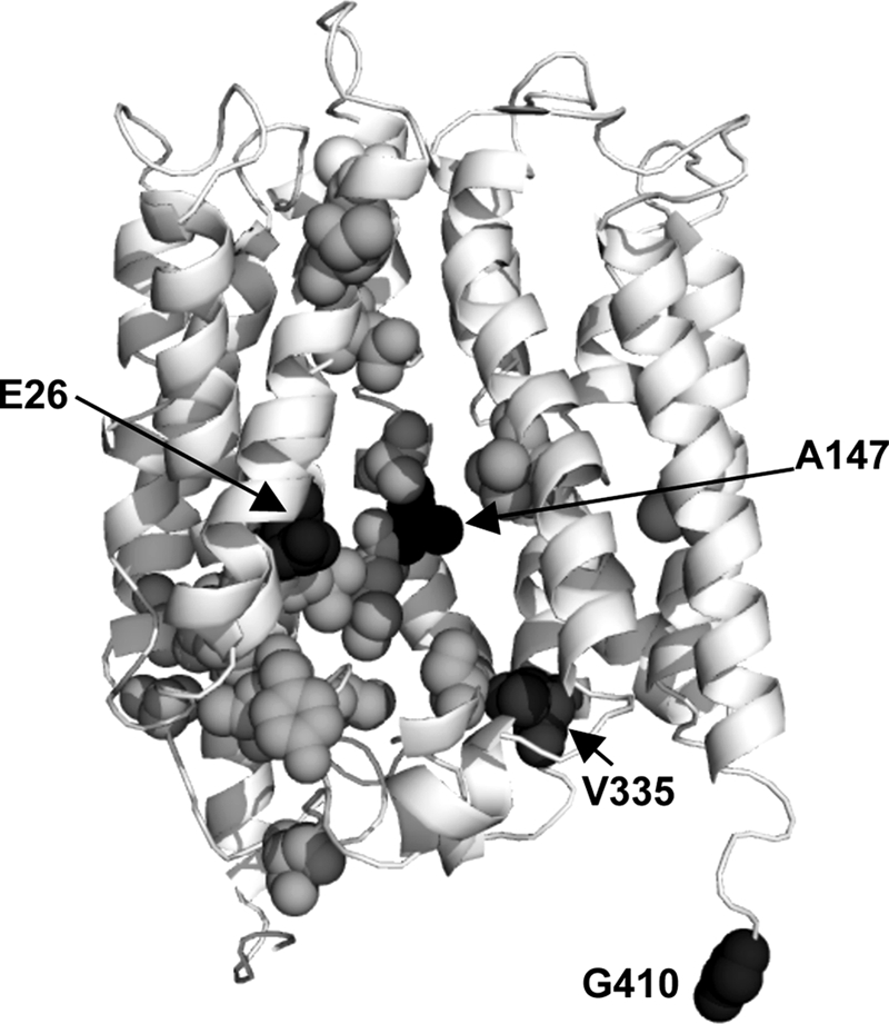
Putative substrate binding residues viewed on the three-dimensional model of MdfA. The ribbon representation of MdfA (11) shows Glu26, and those residues that were studied here at position 147, 335, and 410 (black spheres). The residues shown as gray spheres are putatively involved in substrate recognition by MdfA (6).